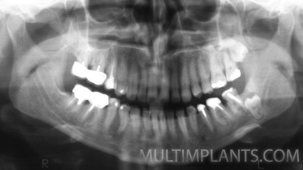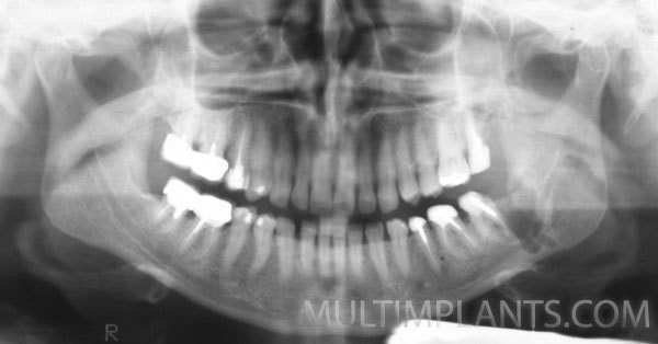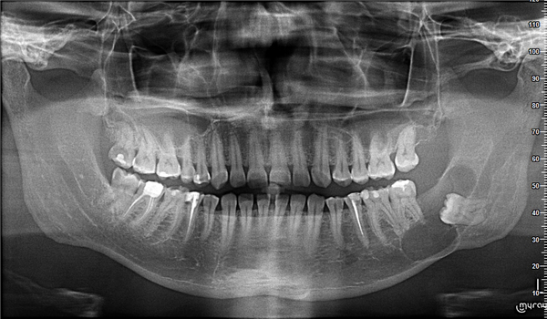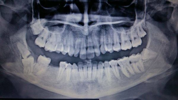


Oral surgery :: IMPACTED /retained/ TEETH
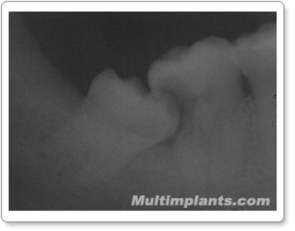
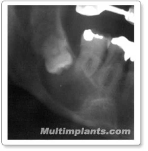
What does a „Impacted“tooth mean? The term derives from the Latin „retentio“- which means hold, i.e. "retained" in the puncture of his teeth. Most often, they strike a third, etc. "canine" teeth and eighths,”wisdom“teeth, etc. This is because these teeth begin to germinate later (after the 13th year for canine teeth and after the 18th - for "wisdom" teeth), when in most cases, the jaws and dental arches are almost shaped and there is simply no place for them. Of course, there are many other reasons that are of concern to specialists and we are not discuss in this patient-friendly material.
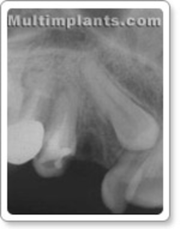
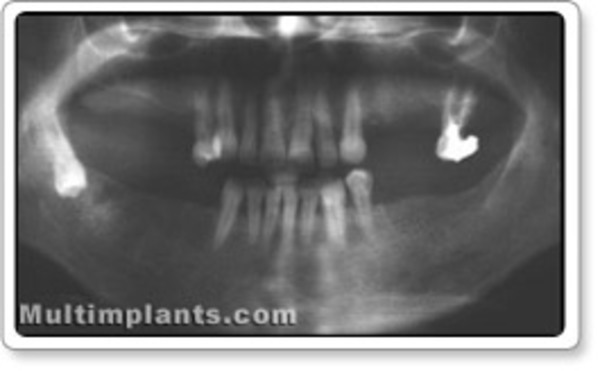
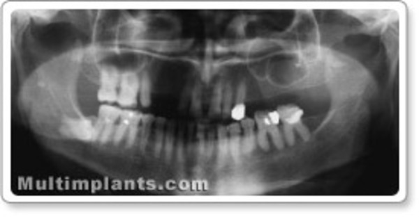
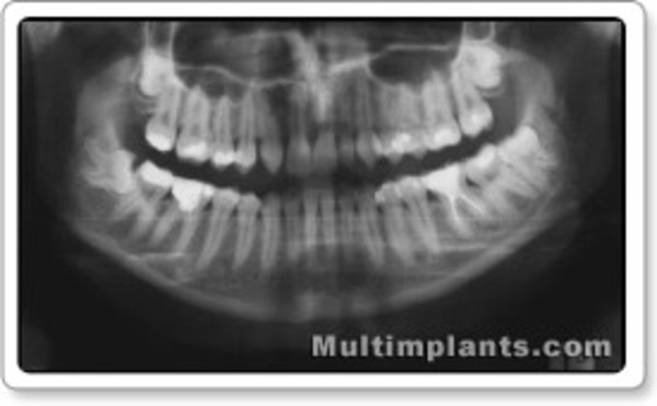
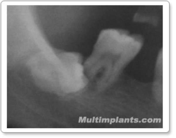
In view of the above, it is easy to understand why these teeth occupy a different position in the bone and adjacent teeth. The lack of space for their emergence does not eliminate the inherent biological potential; they tend to move and do it, but this movement is in the order of microns. This is enough to put pressure on the bone or adjacent teeth due to this potential. Exactly this pressure leads to the destruction of the roots of the adjacent teeth and to the puncture of the bone and mucous membrane, which in turn creates conditions for infection and chronic inflammation in this area, anomalies in the bite and, of course, pain.
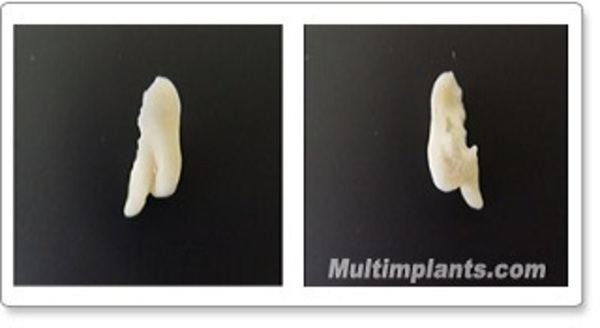
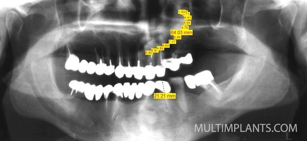
All this requires the extraction / extraction / of such teeth. The task, however, is complex because unforeseen situations arise during the operation itself, which in turn requires extensive experience from the operator. The task is further complicated because, despite perfect operative work, postoperative complications cannot be prevented. To remove the retained tooth, a specific operation perform to remove the bone tissue around the tooth and cut it into as many pieces as necessary. This is a major trauma to the body, and given that there is usually some inflammation at this site and that hundreds of microorganisms live in the oral cavity, it becomes clear why postoperative complications occur, most often in the lower 8-th teeth. The explanation lies in the fact that the anatomy of the jaw in this region is such that in order to reach and remove the tooth, a large amount of bone needs to remove. This is a major trauma to the jaw. In addition, there are tissue lodges, the infection of which is a serious problem in dental practice.
Therefore, extraction of the lower 8-th teeth is the most difficult dental intervention.
For many years, the subject of professional dispute was when to extract the retained teeth-sooner or later. The opinion is now unanimous in favor of early extraction. Motives - still unformed roots, straight roots, lack of infection, lack of adhesions of the tooth with surrounding bone and soft tissues, lack of contact with the mandibular canal, good general condition of the body. However, the usual postoperative complications are pain, swelling, difficulty opening the mouth, difficult and painful swallowing, bleeding.
Less common are complications such as osteomyelitis, abscesses and phlegmons, neuritis and paresis. All this affects the general condition of the body.
Given that retained teeth support chronic inflammation, they often referred to as "focusses". In medicine, the doctrine of so-called "focal infection" has become a separate science. Teeth and many diseases are one of the most common "outbreaks". Disintegrated protein products, which derive from chronic inflammation and specific microorganisms at such sites through a complex mechanism, cause diseases in other organs and systems, most commonly in the heart, kidneys, joints, etc. Therefore, some precautions needed to minimize this type of complication. We give the patient two days before surgery an antibiotic of the penicillin or macrolide group, a non-steroidal anti-inflammatory drug, and an antihistamine drug that lasts for several days after surgery.
A very rare but particularly unpleasant complication is a jaw fracture. Careful and incompetent manipulation with the levers during the intervention, the presence of pathological processes around the tooth such as osteomyelitis, cyst, some types of tumors, etc. The abnormal position of the tooth and its relatively large size compared to the thickness of the jaw is also a reason for this.
In this situation, the doctor may not be guilty of breaking the jaw. In addition, the jaw may break for some time - days, weeks, or months after surgery or even if there is still a tooth in it. This is due to the weakening and to the already anatomically weak spot - the angle of the lower jaw. All of the above is treatable, but requires the patient's prior knowledge and written consent of the risk to which he or she is exposed.
Contact pfhristov@yahoo.com for more details
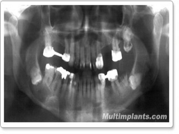
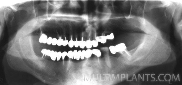
Galley
Check our photo gallery