

Oral implantology :: Hypodontia
The number of teeth is constant with both at temporary and permanent teeth. Sometimes, however, there are deviations from these norms, which we generally divide into three groups:
1. Complete lack of teeth - anodontia
2. Partial lack of teeth or incomplete tooth - hypodontia
3. Ultra-numerous dentition – hyperodontia
We find the incomplete tooth in the literature under the following names: hypodontia / hypodontia /, partial anodontia / anodontia partialis by Damitel /, oligodontia / oligodontia by Bradant /, subtotal anodontia / anodontia subtotalis by Wilner / etc.
We are talking about a hypodontia, when one or more teeth are missing after the normal puncture period and when the tooth enters the occlusion. This anomaly is more a phenomenon of the permanent teeth, from which the 8-th teeth are most often missing, then the fifth lower and rarely the second upper teeth. We come across mainly as a single hypodontia, but there are many symmetrically manifested cases.
There are two types of hypodontia in the clinic:
A / Actual hypodontia (hypodontia vera) when the germ of the unpierced tooth is missing.
B / Apparent hypodontia / pseudohypodontia / when the embryo exists but there is a barrier to its breakthrough / at retained teeth /.
Hypodontia is more common in cultural peoples: Scandinavia - 6%, Europe - 2 - 4%, color - 1% / Lepoivre /
The reasons for hypodontia are various, but the most probable for now are the following: heredity - cases of transmission of hypodontia 2 - 3 generations in a row have been observed; embryo atrophy due to mechanical pressure - at short jaws and pressure from already germinated neighboring teeth, such as biologically active first molars and canine teeth; dental-jaw dystrophies in chronic intoxication - excessive use of alcohol, industrial toxicosis, X-rays, etc .; ectodermal dysplasia - as an element of Crist-Siemens syndrome with the characteristic triad - hypodontia, dyshidrosis and hypotrichosis; in endocrinopathies - disorders of the function of the pituitary and adrenal glands; as involutive reduction of teeth during phylogeny due to changes in diet and diet.
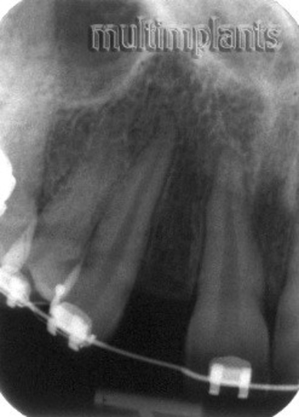
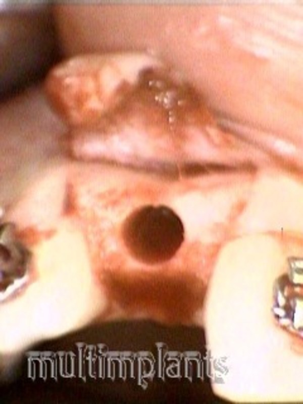
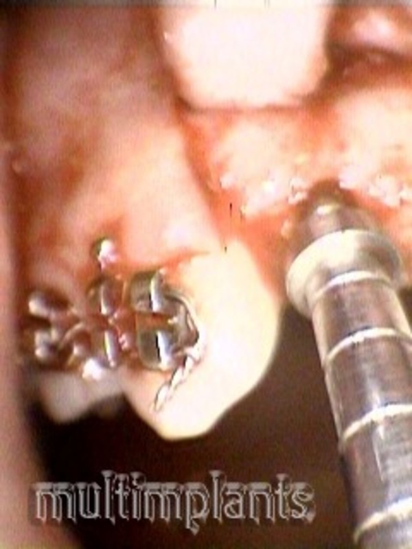
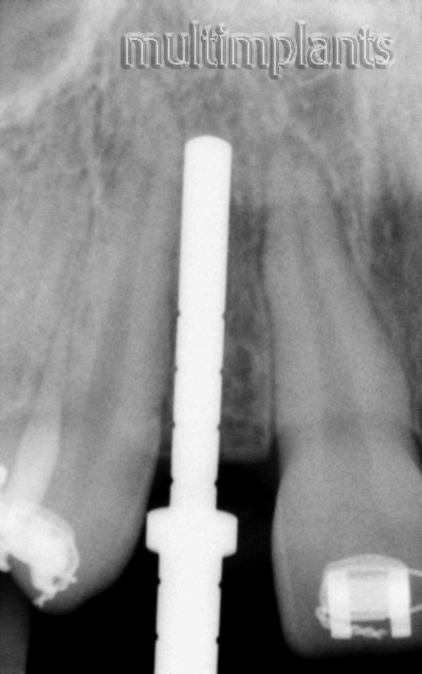
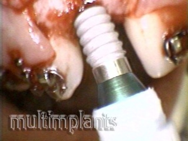
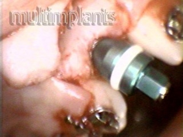
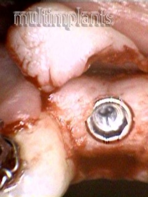
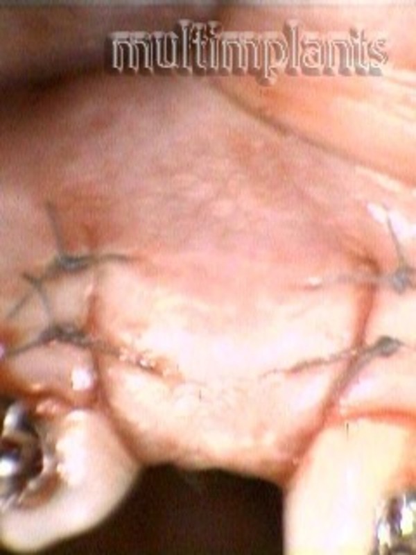
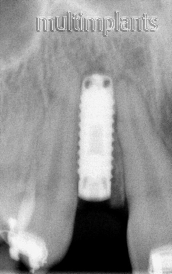
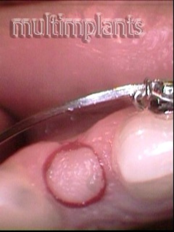
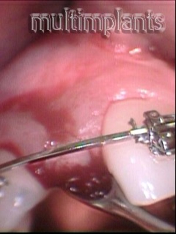
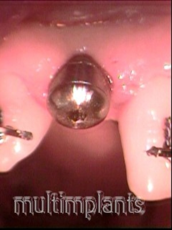
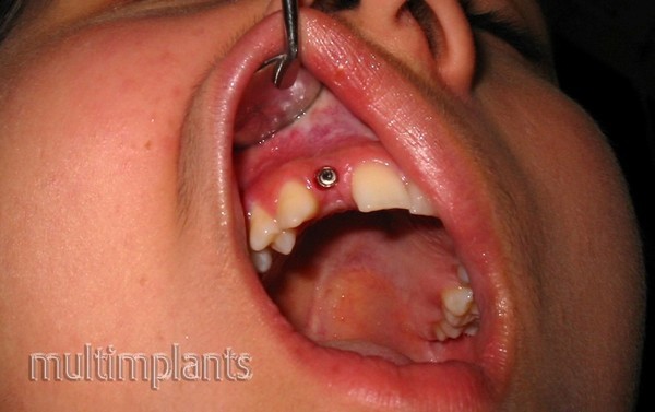
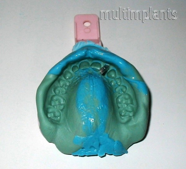
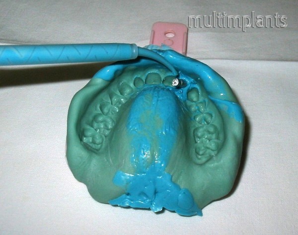
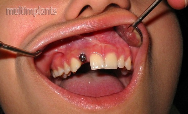
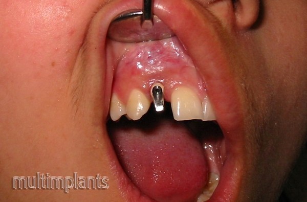
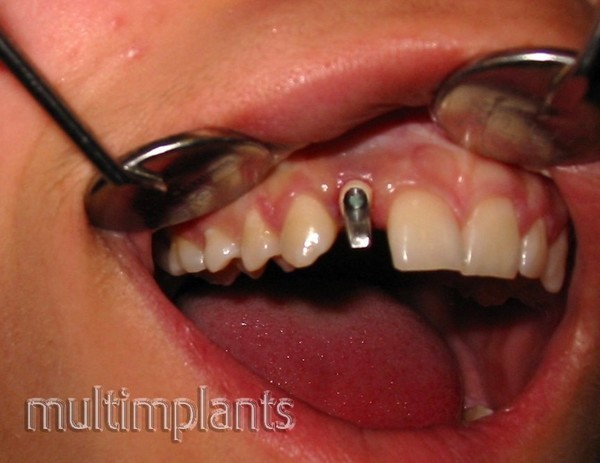
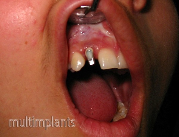
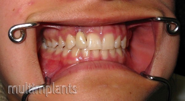
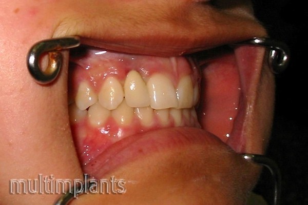
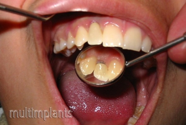
We bring to your attention a case of unilateral hypodontia of the upper right lateral incisor in a 15-year-old girl and the way of treatment.
JM - a 15-year-old girl with no upper right lateral incisor. She treats an anomaly at an orthodontist who directs it for implantation, since the parents do not want on the right 3-th tooth we put metal- ceramics crown, imitating a lateral incisor.
Diagnosis: Hypodontia 12
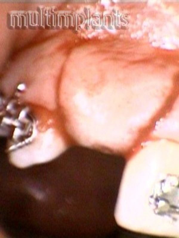
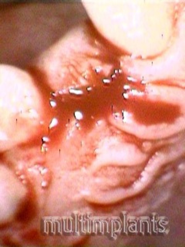
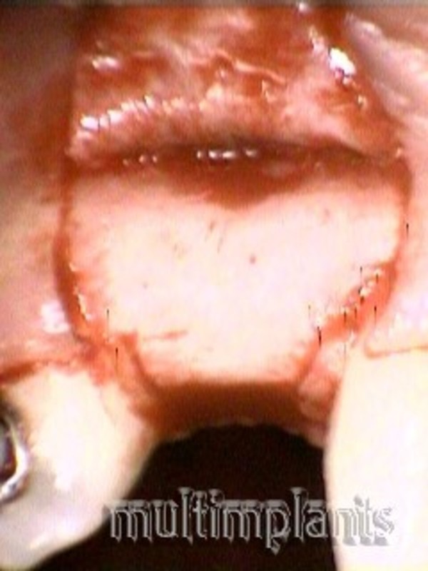
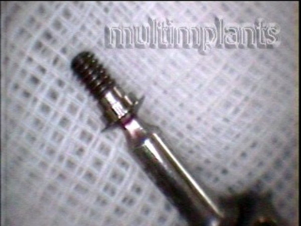
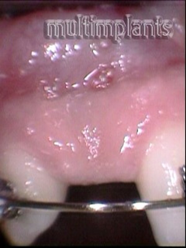
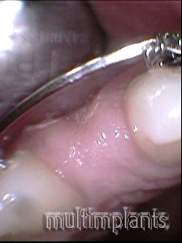
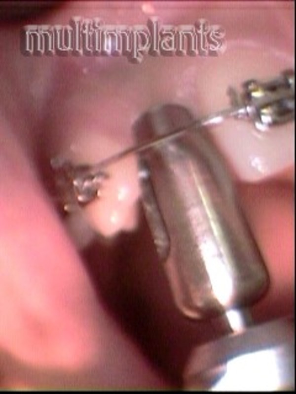
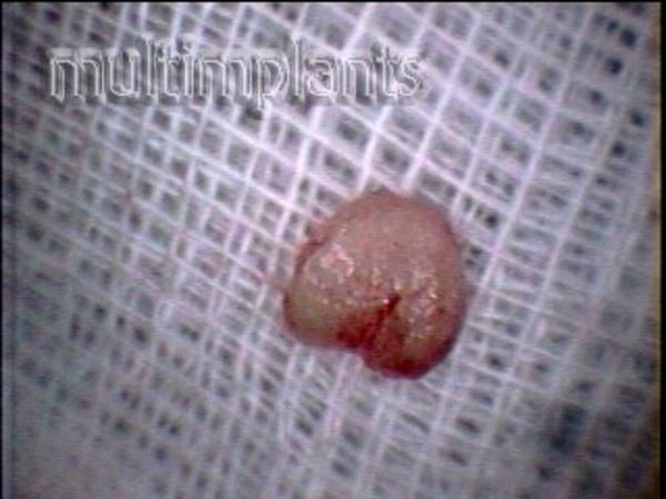
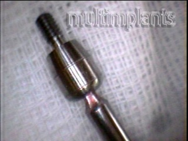
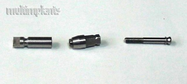
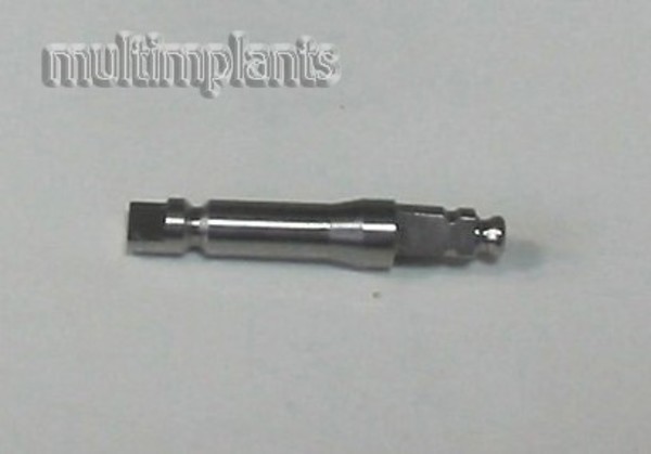
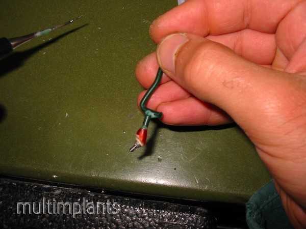
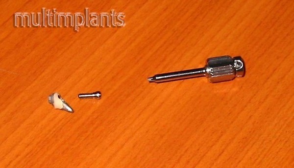
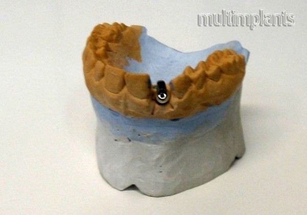
Together with the orthodontist, we decided to use brackets to widen the distance between 11 and 13 teeth by 2 - 3 mm. 6- 6.5 mm. After a few months, we reached this extension and undertook a dental implant T.B.R. ide @ and we envisaged the placement of a metal-ceramic crown on the implant.
Galley
Check our photo gallery